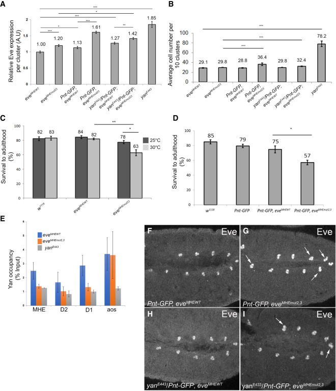
Figure 5.
Loss of robustness in evemut2,3 embryos highlights the importance of ETS2,3-mediated regulation in cardiac cell fate specification. (A) Quantification of average Eve levels per cluster shows that the eveMHEmut2,3 background is sensitized to genetic perturbations that are well buffered against in eveMHEWT embryos. Error bars show SEM. Statistical significance after Bonferroni correction is depicted. (*) P < 0.05; (**) P < 0.01; (***) P < 0.001. (B) The average number of Eve+ cells per 10 clusters in the same genetic backgrounds as in A shows that specification of ectopic Eve+ cells occurs in the eveMHEmut2,3 background, but not in the eveMHEWT control, when the Pnt:Yan ratio is increased. Error bars show SEM. (C) The reduced survival of eveMHEmut2,3 embryos to adulthood was enhanced by temperature stress. (D) Increased pnt dose also decreased eveMHEmut2,3 survival. (E) ChIP-qPCR (chromatin immunoprecipitation [ChIP combined with quantitative PCR [qPCR]) from stage 11 embryos. Signals were normalized to a negative control (see Supplemental Methods). Mutation of the ETS2,3 site reduced occupancy at all three eve CRMs but not at argos (aos). (F–I) Eve expression in representative stage 11 embryos. Examples of clusters with four or more Eve+ cells are indicated with white arrows. Error bars show SEM.

