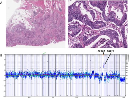
Figure 3.

(A) Moderately differentiated adenocarcinoma of the colon in a 72‐year‐old white man. The tumor invaded through the colon wall and involved numerous pericolonic lymph nodes (pathologic classification T3N2A). The patient rapidly developed stage IV disease. (B) The copy number plot below the histologic images demonstrates extremely high‐level amplification of ERBB2 at 60 copies, associated with lower level coamplification of topoisomerase (DNA) II Alpha (TOP2A) at 7 copies. Using comprehensive genomic profiling, this metastatic colorectal cancer also harbored base substitutions in KRAS (G12D), F‐box/WD repeat‐containing protein 7 (FBXW7) (R479Q), adenomatous polyposis coli (APC) (Q1367*), SRY‐box 9 (SOX9) (D274fs*22), and tumor protein p53 (TP53) (C275W).
