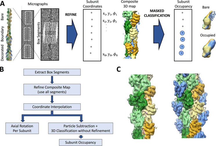
Figure 1.
Identification of cofilin boundaries in cryo-EM micrographs of partially decorated actin. A, graphical representation of the procedure used in this work for analysis of actin filaments heterogeneously decorated by cofilin. An illustrative heterofilament model (left) is built from cofilactin (PDB code 3J0S (10)) and bare actin (PDB code 3J8I (28)) structures. Cofilin is colored blue; actin is colored in shades of green and orange. Adjacent to the model is a representative cryo-EM micrograph of a partially decorated filament with a single boundary between bare (top) and cofilin-decorated (bottom) regions and a close-up of the boundary region. Single-particle cryo-EM refinement and analysis yields the position and orientation of filament subunits within the micrograph, including axial rotations (φ) to be used for subsequent twist analysis. Subsequent masked 3D classification, without coordinate refinement, of individual subunit sites reveals two predominant structural classes, corresponding to bare and cofilin-decorated actin (right). B, flow chart of the procedure depicted in A. C, unsubtracted reconstructions of the bare (left) and cofilactin (right) classes for pyrene-labeled actin, corresponding to the assigned classes illustrated in A.

