Figure 6.

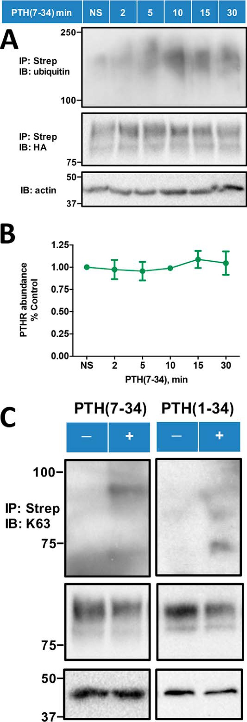
PTH(7–34)-induced receptor ubiquitination. A, PTH(7–34) stimulates receptor ubiquitination in a time-dependent manner. HEK-293S GnTI− cells stably expressing TAP(HA)-PTHR were transiently transfected with Myc-ubiquitin using Lipofectamine 3000. 48 h post-transfection, cells were serum-starved for 2 h and subsequently treated with 10 μm MG132 for 30 min and then challenged with 1 μm PTH(7–34) for the indicated time (0, 2, 5, 10, 15, or 30 min). PTHR was isolated on streptavidin-agarose beads. Ubiquitin was detected with a global anti-ubiquitin antibody that recognizes both Lys48 and Lys63 (P4D1) (top) and PTHR with an anti-HA antibody (middle). β-Actin was used as the loading control (bottom). Molecular weight standards are shown at the left. The illustrated blot is representative of three independent experiments. B, quantification of WT-PTHR expression upon PTH(7–34) treatment. The calculation was performed as described in the legend to Fig. 2C. Results are reported as means ± S.D. (error bars) (n = 3) and indicate that PTHR expression did not change for the first 30 min after challenge with PTH(7–34). C, PTH(7–34) and PTH(1–34) promoted specific Lys63-linked PTHR polyubiquitination. The experimental procedure was similar to that in A, where the protein sample was now detected with a Lys63 linkage-specific polyubiquitin antibody (Table 3) (top) and an anti-HA antibody for receptor expression (middle). β-Actin was used as the loading control (bottom). Positions of molecular weight standards are indicated at the left. Shown is a representative blot from three independent experiments.
