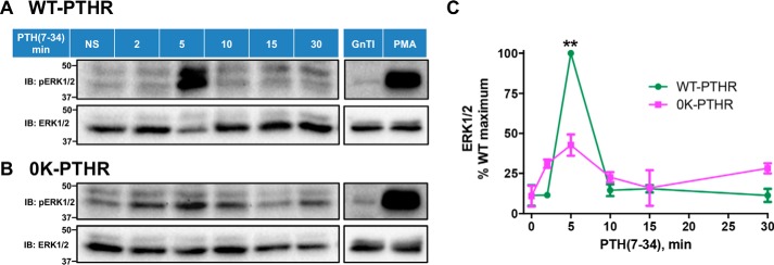
Figure 7.
WT-PTHR and 0K-PTHR mediate distinct patterns of ERK signaling. HEK-293S GnTI− cells stably expressing WT-PTHR (A) or 0K-PTHR (B) were transfected with Myc-ubiquitin in 10-cm dishes. 36 h later, cells were split into six equal aliquots and incubated on 6-cm dishes for 16 h. Cells were serum-starved for 4 h and stimulated with 1 μm PTH(7–34) for the indicated times. 1 μm PMA for 10 min served as a positive control, and native GnTI− cells were transiently transfected with Myc-ubiquitin as a negative control. Cells were immediately washed and lysed as detailed under “Experimental procedures.” Total protein concentrations were measured with BCA. Western blot analysis using equal amounts of total protein from the corresponding cell lysates was performed to detect phosphorylated ERK1/2 (top) and total ERK1/2 (bottom). Positions of molecular weight standards are marked at the left. C, quantitative analysis of ERK signaling. Phospho-ERK signaling was normalized to that of total ERK at each time. Results are means ± S.D. (error bars) of three independent experiments (**, p < 0.01, two-way ANOVA).

