
FIGURE 1.
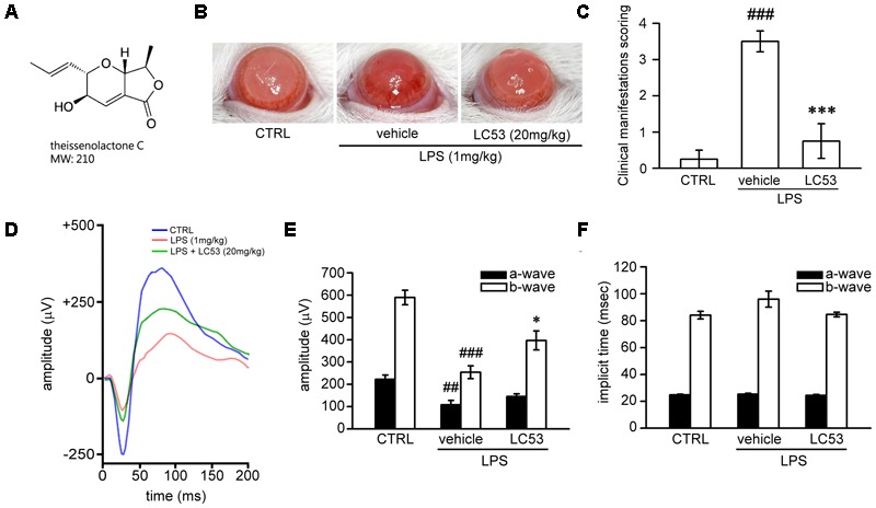
LC53 potentially protected against LPS-induced intraocular inflammation and prevented LPS-injured retinal function. (A) The chemical structure of LC53 (MW = 210). (B) Anterior ocular inflammation was evaluated by microscopic examination in anterior segment 72 h after footpad injection of lipopolysaccharide (LPS, 1 mg/kg). Representative ocular photography was taken at 72 h following LPS-induction in SD rat. (C) Clinical scoring for inflammation level of EIU in the absence (n = 4) or presence of LC53 (20 mg/kg, n = 4) and in control group (n = 4) were determined. (D) Representative scotopic ERG responses from control mice (blue curve), and EIU rat treated with the vehicle control (red curve) or LC53 (green curve) were recorded 1 day after LPS-injection. (E,F) Quantification of the average amplitudes and implicit time (time to peak) from control rat (n = 6), and EIU rat treated with the vehicle control (n = 6) or LC53 (n = 5). The ERG a-waves were negative, and the a-wave amplitudes were presented as the absolute value. The results were presented as the mean ± SEM. CTRL: control. ##p < 0.01, ###p < 0.001 compared with the control group treated with normal saline; ∗p < 0.05, ∗∗∗p < 0.001 compared with EIU group treated with vehicle.