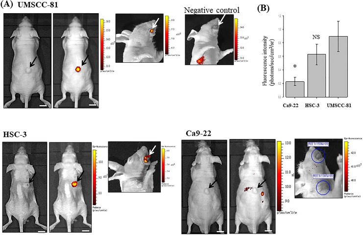
Figure 3.
Near-infrared imaging of OSCC xenografted tumors in mice using the ICG-labeled antipodoplanin antibody and IVIS detection device. A, Brightfield (left panel) and dark field images (right panel, pseudocolor image) of subcutaneous tumors (black arrows) and a dark field image of a tongue tumor (white arrows) approximately 1 week after the injection of 3 OSCC cell lines. UMSCC-81 and HSC-3 subcutaneous tumors with high and moderate podoplanin expression, respectively, are clearly visible, whereas Ca9-22 tumors with weak podoplanin expression are virtually invisible. No significant ICG fluorescent signal was observed in the UMSCC-81 tongue tumor using NIR imaging with an ICG-labeled rat IgG as a negative control (upper right panel). B, Quantitative analysis of NIR fluorescent intensity of UMSCC-81, HSC-3, and Ca9-22 subcutaneous tumors in mice (n = 4). Bar = standard deviation.*P < .05 compared with UMSCC-81 tumors. ICG indicates indocyanine green; IgG, immunoglobulin G; NIR, near-infrared; NS, not significant; OSCC, oral squamous cell carcinoma.

