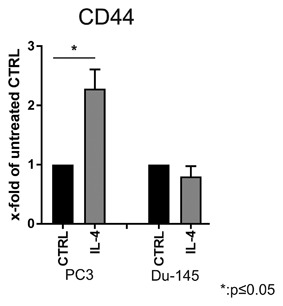
Figure 1.

CD44 expression in PC3 and Du‐145 cell lines after long‐term IL‐4 treatment. Flow cytometry analysis of expression of the surface marker CD44. Before staining, PC3 and Du‐145 cells were treated for 20 passages with 5 ng/mL IL‐4 (n = 3, *P ≤ 0.05)
