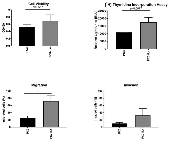
Figure 5.

Increased migration of PC3‐IL4 cells. Functional comparison of PC3 and PC3‐IL4 cells. (A) Cell viability was determined by the EZ4U assay. (B) Cellular proliferation was assessed by measurement of [3H] Thymidine Incorporation. (C) Migration and (D) invasion through Matrigel were assessed by Boyden Chamber assays (n = 3, *P ≤ 0.05)
