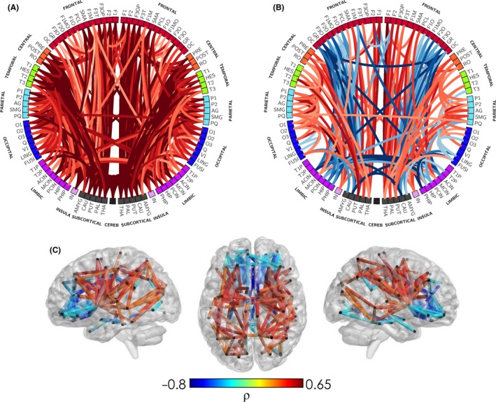
Figure 3.

Network representation of developing microstructure. The correlation of corrected age at MRI with structural connections weighted by NODDI intracellular volume fraction index (or neurite density index, NDI), in a population of 65 infants scanned between 25 and 45 weeks of postmenstrual age (PMA). In panel (A) NDI parameters increase with age for most white matter connections, as would be expected. However, when assessed in relative terms (%) of the total connectivity in each subject (panel B and C), it is possible to separate which connections are developing at a relative faster or slower pace. This clarifies the expected heterochronicity in the early development of brain connectivity, with a general trend of connections between somatosensory, central, subcortical and temporal areas to show faster development (red) than frontolimbic and interhemispheric connections (blue). Adapted from Batalle et al. (2017) [Colour figure can be viewed at http://wileyonlinelibrary.com]
