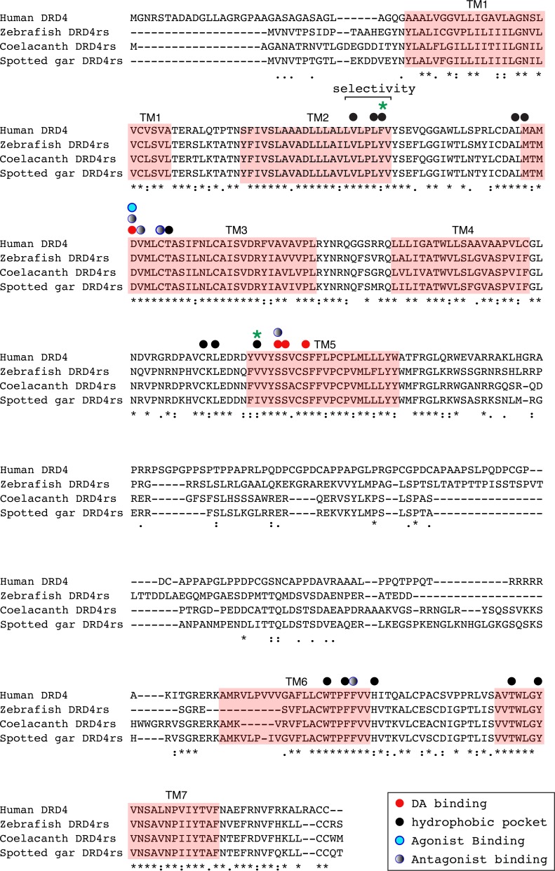
Figure 8. Alignment of the human dopamine receptor 4 (DRD4) with zebrafish (Danio rerio), coelacanth (Latimeria chalumnae) and spotted gar (Lepisosteus oculatus) dopamine receptor 4rs (DRD4rs).
Shaded regions denote transmembrane domains according to UniProt. Dopamine binding sites (red dots) were determined by site directed mutagenesis (Cummings et al., 2010) and homology to DRD2. Antagonist binding sites and hydrophobic pocket-including selectivity region-were obtained from mutagenesis studies (Cummings et al., 2010) and from the crystal structure of the receptor coupled to the antagonist nemonapride (Wang et al., 2017). Non-conserved amino acids in the nemonapride binding pocket are labeled with green asterisks. Binding sites for the selective agonist UCSF924 are also shown (light blue dot).

