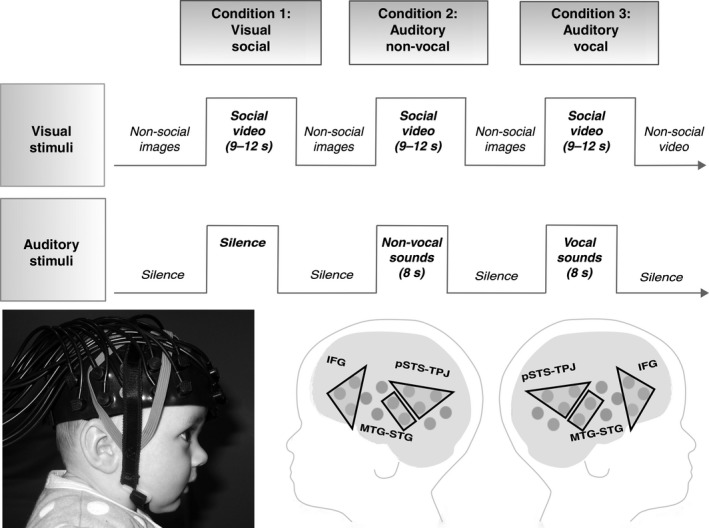
Figure 1.

Upper panel: stimulus presentation. Lower panel: An infant wearing the fNIRS headgear (left) and the three ROIs projected on a schematic of the fNIRS measurement channels (right) – inferior frontal gyrus – IFG, posterior portions of the superior temporal sulcus region – temporoparietal junction – pSTS‐TPJ and anterior portions of the middle temporal gyrus – superior temporal gyrus – aMTG‐STG. Parental consent was obtained for the publication of this photo.
