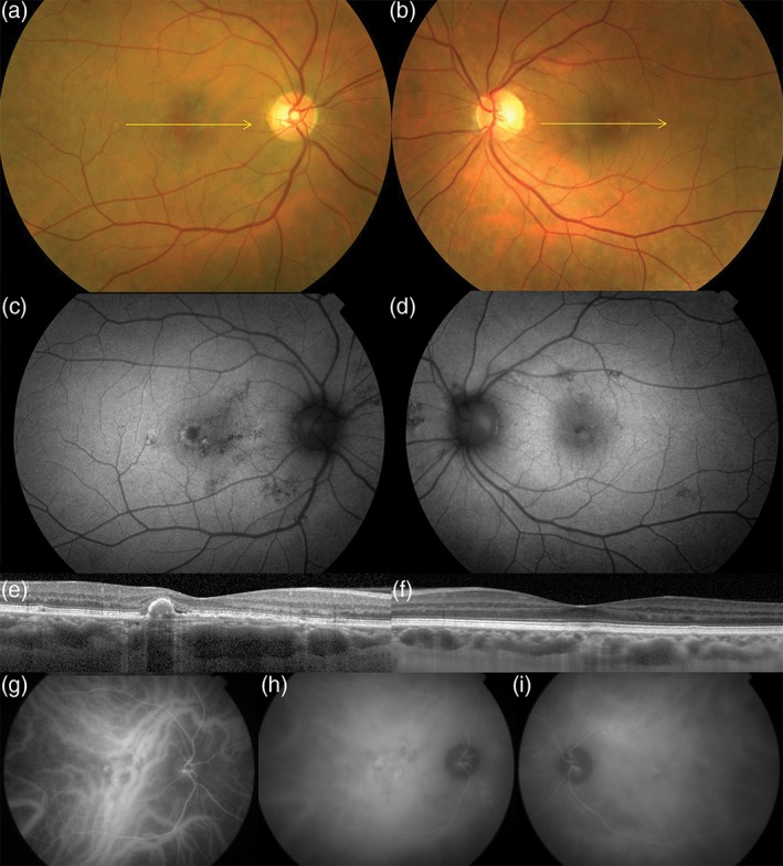
Figure 4.

Aneurysmal type 1 neovascularization (polypoidal choroidal vasculopathy) in a 57‐year‐old white female. Colour photographs of the right (a) and left (b) eyes show absent drusen in the posterior poles. Fundus autofluorescence of the right (c) and left (d) eyes show hyper‐ and hypoautofluorescent changes in the macula and nasal to the optic nerves. Spectral domain optical coherence tomography of the right (e) and left (f) eyes. A shallow irregular pigment epithelial detachment is present in the right eye (e) with a peaked component, consistent with a type 1 neovascularization and an aneurysmal (polypoidal) lesion, respectively. Dilated Haller's layer veins are present in both eyes, especially so in the right eye (f) where, at the site of neovascularization, the inner choroid is attenuated. Early phase indocyanine green angiography of the right eye (g) shows pathologically dilated choroidal veins traversing the fovea with subtle hyperfluorescence of an overlying type 1 neovascular lesion (branching vascular network). A more intensely hyperfluorescent aneurysmal (polypoidal) lesion is seen at the temporal edge of the type 1 neovascular lesion. A hyperfluorescent plaque corresponding to type 1 neovascular lesion is seen in the mid‐phase (h). Areas of choroidal hyperpermeability are present nasal to the optic nerve in the right eye (h) and in the superotemporal macula of the left eye (i).
