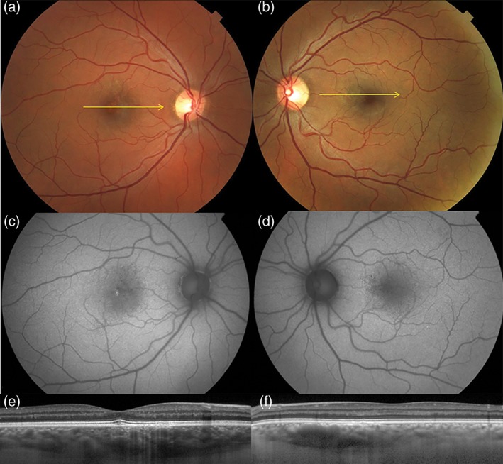
Figure 5.

Pachychoroid pigment epitheliopathy in a 37‐year‐old white male. Colour photographs of the right (a) and left (b) eyes show absent drusen and reduced fundus tessellation. Fundus autofluorescence of the right (c) and left (d) eyes show non‐specific pigment epithelial changes, including a small focus of hyperautofluorescence at the right fovea (c). Spectral domain optical coherence tomography of the right (e) and left (f) eyes shows thick choroids in both eyes. In the left eye (f), a region of extrafoveal maximal choroidal thickness is attributable to Haller's layer vessel dilatation, anterior to which the inner choroid is attenuated.
