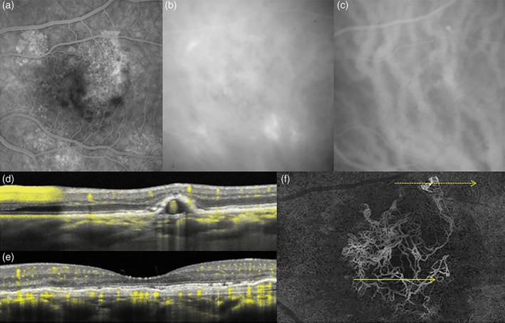
Figure 6.

Fluorescein angiography (FA), indocyanine green angiography (ICGA) and optical coherence tomography (OCT) angiography of the left eye in 59‐year‐old white male with aneurysmal type 1 neovascularization (polypoidal disease). Non‐specific hyperfluorescence is seen on FA (a) while ICGA (b, c) shows dilated Haller's layer veins, choroidal hyperpermeability, and a focal area of hyperfluorescence (arrow). Dense raster cross‐sectional OCT (d, e) shows a shallow irregular pigment epithelial detachment (PED) in the central macula and a peaked extrafoveal PED, consistent with aneurysmal type 1 neovascularization (polypoidal disease with a branching vascular network). The angiographic signal overlay (yellow pixels) confirms flow within the shallow PED (e) and the aneurysmal lesion (g). En face swept source OCT angiography (f) segmented immediately anterior to Bruch's membrane reveals the morphology of the type 1 neovascular network, the distant location of the aneurysmal lesion and its feeding vessels originating from the neovascular tissue. The direction and location of the dotted and yellow arrows correspond to (d) (upper arrow) and (e) (lower arrow) cross‐sectional scans, respectively.
