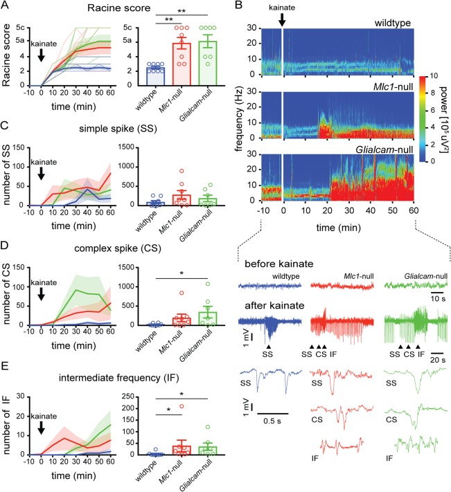
Figure 4.

Lowered seizure threshold in MLC mice. (A) Behavioral scoring of seizure activity over time in wild‐type (blue) and MLC mice (Mlc1‐null: red; Glialcam‐null: green; thin lines show individual mouse responses; thick lines show averages) following kainate injection (10mg/kg intraperitoneally). For each 10‐minute interval, the highest level of epileptic activity was scored using the modified Racine seizure scale. The bar graph shows maximum Racine score during 60‐minute trial (open circles indicate individual mice, bars show average). MLC mice reached significantly higher behavioral seizure scores (wildtype: n = 8; Mlc1‐null: n = 8; Glialcam‐null: n = 7; p = 0.0014; wildtype vs Mlc1‐null, p = 0.0060; wildtype vs Glialcam‐null, p = 0.0023). (B) Top: Morlet‐wavelet ECoG spectra from kainate‐injected mice. Bottom: ECoG traces showing simple spike waves (SS), complex spike waves (CS) and runs of intermediate frequency (IF) discharges (expanded below). (C–E) Temporal progression of kainate‐induced SS, CS, and IF events, respectively. Bar graphs show total number of SS, CS, and IF events in 60‐minute trial. A significantly higher number of IF discharges was observed in MLC mice (wildtype: 4.1 ± 2.9 discharges/h; Mlc1‐null: 39.0 ± 24.8; Glialcam‐null: 35.4 ± 15.8; p = 0.0101; wildtype vs Mlc1‐null, p = 0.028; wildtype vs Glialcam‐null, p = 0.012). Error bars and shaded regions indicate SEM. *p < 0.05; **p < 0.01; ***p < 0.001. ECoG = electrocorticogram; MLC = megalencephalic leukoencephalopathy with subcortical cysts.
