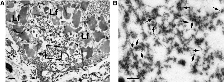
FIGURE 3.
Immunoelectron microscopy with Perry syndrome. (A) A neuron from the subthalamic nucleus has a cytoplasmic inclusion (arrow) surrounded by lipofuscin (Lf). Boxed area is enlarged in (B). (B) The inclusion contains granule-associated filaments immunopositive for phospho-TDP-43 antibody (arrows point to gold particles). Scale bars = 1 μm in (A); 200 nm in (B).

