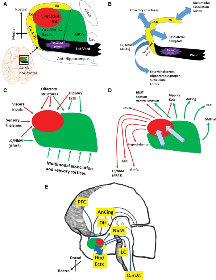
FIGURE 4.
The amygdala is an approximately 1.5-cubic-centimeter amalgamation of cell groups, with complex connections that include regions of the brain that are vulnerable to neurodegenerative disease pathologies. (A) A schematically rendered axial/horizontal oriented profile of the amygdala to depict some surrounding structures, as well as the 3 main regions of the amygdala: the basolateral complex (green), the anteriomedial area (red), and the medial cortex-like zones (yellow), roughly following de Olmos et al (94); this is not an accurate depiction because not all of these structures are in the exact same plane. (B) The medial cortex-like zone of the amygdala is the least well understood in terms of connectivity in human brains, but has strong reciprocal relationships with the hippocampal formation, entorhinal cortex, and olfactory structures. (C, D) Inputs (C) and outputs (D) of the basolateral complex and anteriomedial region are better understood from prior studies performed in rodents and nonhuman primates. Within the amygdala, information flows from the basolateral and accessory nuclei, toward the anteromedial nuclei, from which derives the main amygdalar output to the diencephalon and brainstem. (E) Panel depicts a right hemibrain to show some of the many brain regions that are directly connected with the amygdala and have been strongly implicated in the earliest phases of various neurodegenerative diseases. More information on amygdala connectivity may be found in (93, 95, 96, 100, 101, 114). Acc. Bas.n., accessory basal nucleus; ag, ambient gyrus; AnCing, anterior cingulate cortex; ARAS, ascending reticular activating system; Bas.n., basal nucleus; Bsl. Forbr., basal forebrain, including magnocellular neurons (nucleus basalis); BNST, bed nucleus of the stria terminalis; Cau, tail of the caudate; Cent.Med.n.g., centromedian nuclear group; Claus, claustrum; Co.n., cortical nucleus; Co.A.Tr., cortico-amygdaloid transition area; cs, collateral sulcus; DMThal, dorsomedial thalamus; d.m.V., dorsal motor nucleus of the vagus nerve; Ectx, Entorhinal cortex; ers, endorhinal sulcus; fg, fusiform gyrus; Ins, insula; Lat.n., lateral nucleus; LC, locus coeruleus; NbM, nucleus basalis of Meynert; PAG, periaqueductal grey matter; PFC, prefrontal cortex; phg, parahippocampal gyrus; sg, semilunar gyrus; Str., striatum/ventral extension of putamen; TEctx, transentorhinal cortex.

