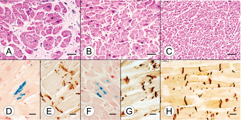
FIGURE 1.
Cardiac pathology in juvenile compound heterozygous and homozygous Friedreich ataxia (FA). (A, D, E) Compound heterozygous FA case 1; boy, 11; (B, F, G) homozygous FA case 1; boy, 10; (C, H) normal control; boy, 9. (A) The cross section of the ventricular septum (VS) in the compound heterozygote shows cardiomyocyte enlargement, excessive lobulation of fibers, and endomysial fibrosis. (B) The comparable section of VS in the homozygous FA patient is similar though endomysial fibrosis is less prominent. (C) The section of the VS from an age-matched control reveals the great abundance of small normal cardiomyocytes and a delicate endomysium. (D, F) Longitudinal sections of the VS in the compound heterozygous FA case 1 (D) and the age-matched homozygous FA case 1 (F) reveal iron-containing inclusions. (E, G) N-cadherin immunohistochemistry shows large disrupted intercalated discs (ICD) in both the compound heterozygous FA case 1 (E) and the age-matched homozygous FA case 1 (G). (H) N-cadherin immunohistochemistry of the normal VS shows small ICD that are aligned in register across adjacent heart fibers. The ICD also display a lesser degree of undulation than in FA (E, G). (A–C) Hematoxylin and eosin; (D, F) Perls's stain for iron; (E, G, H) immunohistochemistry for N-cadherin. Scale bars: (A–C), 50 μm; (D–H), 20 μm.

