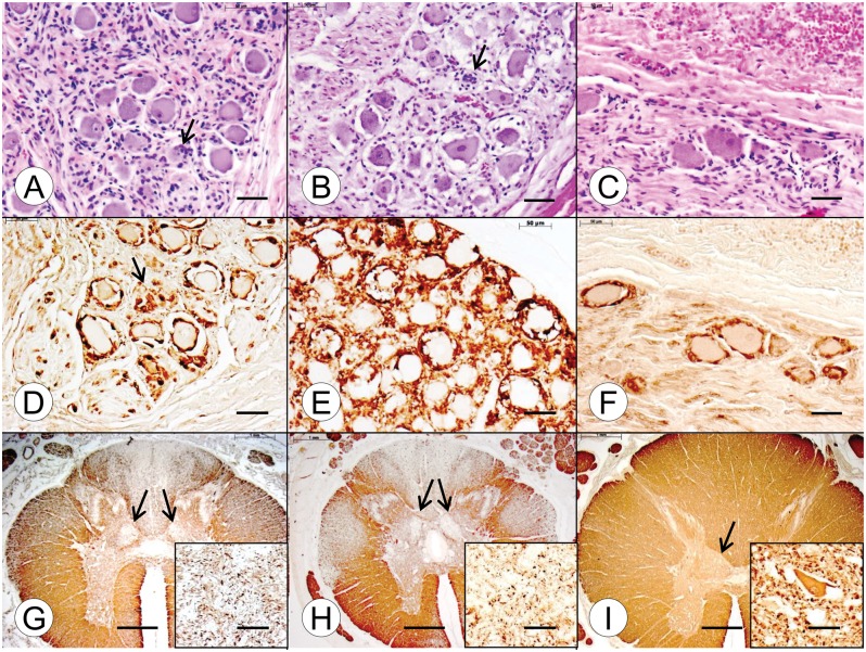
FIGURE 3.
Dorsal root ganglia (DRG) and thoracic spinal cord in juvenile compound heterozygous and homozygous Friedreich ataxia (FA). (A, D, G) Compound heterozygous FA case 1; boy, 11; (B, E, H) homozygous FA case 1; boy, 10; (C, F, I) normal control; boy, 9. (A–C) DRG in the compound heterozygous (A) and homozygous FA (B) cases show hypercellularity and residual nodules (arrows). The overall neuronal size in the FA cases is smaller than normal (C). (D–F) Prominent S100 reaction product is present in satellite cells of the DRG in the compound heterozygous FA case 1 (D) and homozygous FA case 1 (E). The arrow in (D) indicates an S100-reactive residual nodule. In the case of homozygous FA (E), S100 immunohistochemistry suggests an abnormal abundance of satellite cells, compared to the normal control (F). (G–I) The cross-sectional area of the thoracic spinal cord in compound heterozygous (G) and homozygous FA (H) is smaller than normal (I). In the compound heterozygous FA case (G) and the homozygous FA case (H), depletion of myelinated fibers in the dorsal columns is comparable. Fiber loss in the dorsal spinocerebellar and lateral corticospinal tracts of the compound heterozygote (G) is less prominent than in the homozygous case (H). The myelin basic protein (MBP) stains also show fiber depletion in the dorsal nuclei in compound heterozygous FA (G, arrows) and homozygous FA (H, arrows). In the normal spinal cord, the dorsal nucleus is distinctly visible (I, arrow). (G–I, insets) Immunohistochemistry for class-III-β-tubulin reveals total loss of large neurons in the compound heterozygous (G, inset) and homozygous (H, inset) FA cases. The class-III-β-tubulin stain in the normal control (I, inset) reveals a large normal neuron with a maximum diameter of approximately 50 μm. (A–C) Hematoxylin and eosin; (D–F) immunohistochemistry for S100; (G–I) immunohistochemistry for MBP; (G–I, insets) immunohistochemistry for class-III-β-tubulin. Scale bars: (A–F), 50 μm; (G–I), 1 mm; (G–I, insets), 50 μm.

