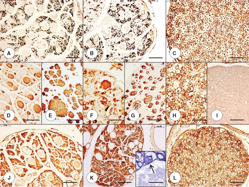
FIGURE 5.
Dorsal roots (DR) in juvenile compound heterozygous and homozygous Friedreich ataxia (FA). (A, D, E, J) Compound heterozygous FA case 1; boy 11; (B, F, G, K) homozygous FA case 1; boy 10; (C, H, I, L) normal control; boy, 9. (A–C) The axon stain shows negative images of expansions within the substance of the DR in compound heterozygous case 1 (A) and homozygous FA case 1 (B). The organized distribution of large and small axons of the normal DR is absent (C). (D–I) The expansions in the DR yield reaction product with anti-S100 in compound heterozygous FA case 1 (D) and homozygous FA case 1 (F). The glial fibrillary acidic protein (GFAP) stain yields similar results (E, G). In the normal control case (H, I), S100 reaction product shows delicate rims around large and small DR axons (H) but no GFAP (I). The DR in compound heterozygous FA (D) and homozygous FA (F) show a deficit of S100 in regions occupied by axons (A, B). (J–L) The expansions in DR are negative for laminin but are surrounded by a delicate layer of reaction product in compound heterozygous FA (J) and homozygous FA (K). The normal DR (L) reveal a honeycomb-like pattern of laminin reaction product around large axon and islands of small axons. This pattern is absent from the DR in compound heterozygous FA (J) and homozygous FA (K). (A-C) Immunohistochemistry for phosphorylated neurofilament protein; (D, F, H) immunohistochemistry for S100; (E, G, I) immunohistochemistry for GFAP; (J–L) immunohistochemistry for laminin; (K, inset) resin-embedded section stained with Toluidine blue. Scale bars: (A–L), 50 μm; (K, inset), 20 μm.

