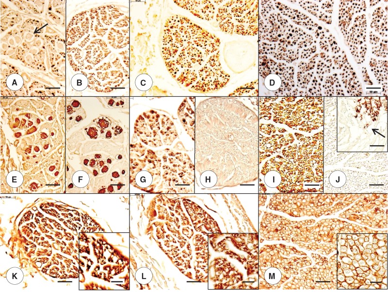
FIGURE 6.
Dorsal roots (DR) in adult compound heterozygous and homozygous Friedreich ataxia (FA). (A, B, E, F, K) Compound heterozygous FA case 2; man, 28; (C, G, H, L) homozygous FA case 2; woman, 25; (D, I, J, M) normal control; woman, 38. (A–D) The DR of the compound heterozygous (B) and the homozygous (C) FA cases indicate a relative lack of large axons. In some DR of the compound heterozygous FA case 2 (A, arrow), the stain reveals similar expansions as illustrated in Figures 5A and B. (E–J) The DR expansions in the compound heterozygous case are reactive with antibodies to S100 (E) and glial fibrillary acidic protein (GFAP) (F). DR in the homozygous FA case show a deficit in S100 reaction product (G) but are GFAP-negative (H). The normal DR in the control specimen displays more abundant S100 reaction product (I) and is GFAP-negative (J). GFAP immunoreactivity attributable to the central nervous system ends at the DR entry zone (J, inset, arrow). (K–M) The DR of the compound heterozygous FA case 2 (K) and the homozygous FA case 2 (L) show compact laminin reaction product around the voids of thin axons (K, L, insets). The normal DR (M) displays the orderly honeycomb-like reaction product of laminin (M and M, inset). (A–D) Immunohistochemistry for phosphorylated neurofilament protein; (E, G, I) immunohistochemistry for S100; (F, H, J) immunohistochemistry for GFAP; (K–M and insets) immunohistochemistry for laminin. Scale bars: (A–M), 50 μm; (J–M, insets), 20 μm.

