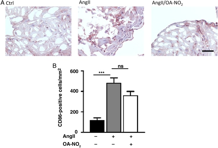
Figure 5.
Number of CD86-positive cells in atrial tissue. (A) Representative figures of an immunohistological staining of atrial sections by using an anti-CD86-specific antibody. (B) Quantification of CD86-positive cells in atrial tissue reveal a significant higher amount of these cells in AngII (+)/OA-NO2 (−) animals (n = 5/4/4). Furthermore, this was not significantly influenced by a parallel treatment with OA-NO2. For the staining of paraffin-embedded murine atrial sections with CD86 (1 : 500, rabbit IgG, NB110–55488, Novus Biologicals, USA), a standard protocol was used. For quantification, CD86-positive cells were counted in atrial sections. Scale bar = 50 µm. ***P < 0.001.

