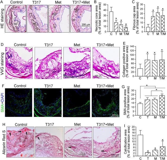
Figure 2.

Co‐treatment of metformin and T317 maintains structural integrity of aortic wall and inhibits calcium deposits in aortic lesion areas. The aortic root cross sections were assessed by the following assays: (A–C) Haematoxylin and eosin staining (A) followed by quantitative analysis of necrotic core area (B) and fibrous cap area (C). nc: necrotic cores marked by a black dashed line; fc: fibrous cap marked by a blue dashed line; (D, E) VVG staining (D) followed by quantitative analysis of collagen content (E); (F, G) immunofluorescent staining for expression of SMA (F) with quantification of SMA positive areas (G); (H, I) Alizarin Red S staining for calcification (H, indicated by black arrows) and quantification of calcification positive areas (I). *P < 0.05, significantly different from control or as indicated (n = 15). C, control; T, T317; M, metformin; T/M, T317 plus metformin.
