Figure 5.
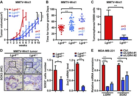
Lgr4 loss impairs breast CSCs. A) The volume of primary tumors until 10 wk from initial tumor detection by palpation in Wnt1-Lgr4+/+ (n = 7), -Lgr4+/ – (n = 9), and -Lgr4−/− (n = 4) mice. B) The time from initial detection to tumor reaching 1.5 cm diameter in MMTV-Wnt1 mice of indicated Lgr4 genotypes. Wnt1-Lgr4+/+ (n = 21), -Lgr4+/− (n = 27), and -Lgr4−/− (n = 6). C) Primary mammary tumor cells from indicated mice were plated under nonadherent conditions for 18 d, and resulting tumorspheres were quantitated (n = 3). D) The protein expression of SOX2 was determined by immunohistochemistry (left) in Wnt1-Lgr4+/+and -Lgr4−/−mammary tumors. Scale bars, 50 µm (top); 20 μm (bottom). Graphs show quantitation of SOX2+ cells (middle) and the SOX2 mRNA level determined by quantitative PCR (right). E) LGR4 and SOX2 mRNA expression in MDA-MB-231 expressing control (ctrl) or indicated siRNA. Error bars are means ± sd. * P < 0.05, **P < 0.01, ***P < 0.001.

