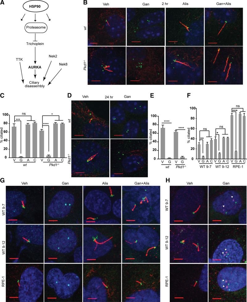
Figure 1.
HSP90 is required for in vitro maintenance of cilia. A) Selected subset of defined proteins that influence ciliary dynamics or structural integrity, and are HSP90 clients or regulated by HSP90 inhibition. B–E) Representative immunofluorescence (IF) image and graph with frequency of ciliated WT or Pkd1−/− murine renal epithelial cells at 2 h (B, C) or 24 h (D, E) after treatment with vehicle (V), ganetespib (G), or alisertib (A) to inhibit AURKA, or combination (C) of alisertib and ganetespib. On IF, acetylated α-tubulin (red); γ-tubulin (green); DAPI (blue). Scale bars, 5 μm; original magnification, ×240. F, G) Analysis as in B and C for human PKD1-mutant WT9-7 and WT9-12 cells or for hTERT-RPE1 (RPE1) cells, 2 h after drug treatment. H) IF for phospho-T288 (activated) AURKA (magenta, indicated by white arrowheads) at ciliary basal body (γ-tubulin, green) with acetylated α-tubulin (red); images of ph-AURKA and basal body are offset. No antibodies currently available for phospho-T288 are reactive with mouse AURKA. Scale bars, 5 μm. Data are expressed as means ± sem; n/s, not significant. IF experiments were performed in triplicate; ≤750 cells were scored per genotype. *P ≤ 0.05, ***P ≤ 0.001, ****P = 0.0001.

