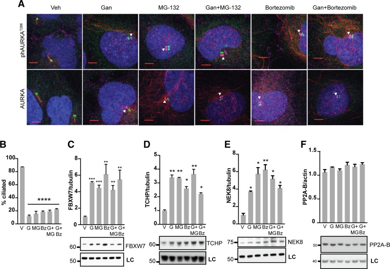
Figure 2.
Mechanism of ganetespib regulation of AURKA activation in ciliary disassembly. A) Immunofluorescence analysis of hTERT-RPE1 cells treated for 2 h with vehicle (Veh), ganetespib (Gan), and/or proteasomal inhibitors MG-132 and bortezomib. DAPI (blue), γ-tubulin (green), acetylated α-tubulin (red), activated ph-T288-AURKA (top row, magenta) or total AURKA (bottom row, magenta). Scale bars, 5 μm; original magnification, ×240. Images of ph-AURKA or total AURKA are offset from basal body. B) Frequency of ciliated cells in populations of cells 2 h after treatment with vehicle (V), ganetespib (G), and/or MG-132 (MG) and bortezomib (Bz). C–F) Normalized quantification and representative data for Western blots showing expression of FBXW7 (C), TCHP (D), NEK8 (E), or PP2A-B (F), 2 h after treatment with indicated drugs. LC, loading control. *P ≤ 0.05, **P ≤ 0.01, ***P ≤ 0.001.

