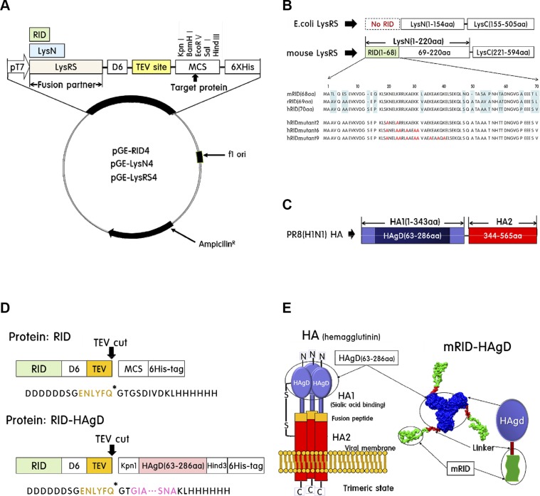
Figure 1.
Construction of the pGE-RID4 vector and HAgD viral protein. A) Schematic illustration of the pGE-RID (4) vector used for the expression of fusion proteins in E. coli. B) Sequence homology of RIDs of LysRS from humans, mice, and rabbits. E. coli LysRS has no RID sequence. C) The globular head domain of HA consists of aa 63–286 in the HA1 subunit. D) Modular representation of the fusion protein. By the TEV protease, the fusion protein is cleaved into an RID and HAgD carrying a 6xHis tag. E) Left: schematic illustration of trimeric HA composed of the HA1 globular domain (containing HAgD) and HA2 of mostly α-helical conformation. Right: structure modeling of mRID-HAgD.

