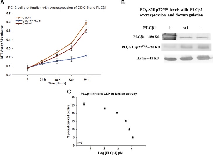
Figure 4.
PLCβ1 inhibits CDK16 activity. A) Increased PC12 cell proliferation with CDK16 overexpression and its reversal with increased PLCβ1 expression; n = 6; control vs. PLCβ1 + CDK16: t = 14.023, P ≤ 0.001; CDK16 + PLCβ1 vs. CDK16: t = 3.887, P = 0.009. B) Western blot of SK-N-SH cell showing the elimination of inactive PO4-Ser10- p27Kip1 when PLCβ1 is overexpressed (n = 6), or slightly down-regulated [i.e., 12-h treatment with siRNA (PLCβ1)] as shown in the third lane. We note that here we overexpressed yellow fluorescent protein–PLCβ (160 kDa) to visually monitor the level of overexpression; this construct has slightly reduced mobility compared with that of the wild-type protein (160 vs. 140 kDa). C) FRET-based commercial assay showing the relative phosphorylation of ∼40 nM CDK16 with increasing PLCβ1 where data is presented. The PLCβ1 concentration is presented in picomolars in logarithmic form so that the full range of PLCβ1 concentrations can be readily viewed (P ≤ 0.001; f = 88.441).

