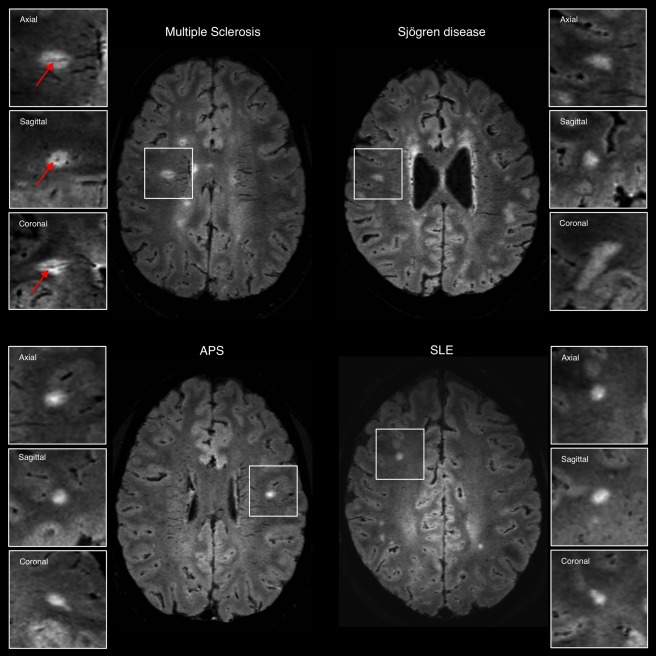
Figure 2.

Representative axial 3T FLAIR* images from individuals with relapsing–remitting multiple sclerosis (MS; 27‐year‐old woman), Sjögren disease (46‐year‐old woman), antiphospholipid antibody syndrome (APS; 37‐year‐old man), and systemic lupus erythematosus (SLE; 38‐year‐old woman). The central vein sign (arrows) is present in the majority of MS lesions but is not typical of white matter lesions in inflammatory vasculopathies. Boxes show magnified views of lesions in the 3 orthogonal planes for central vein assessment. [Color figure can be viewed at http://www.annalsofneurology.org]
