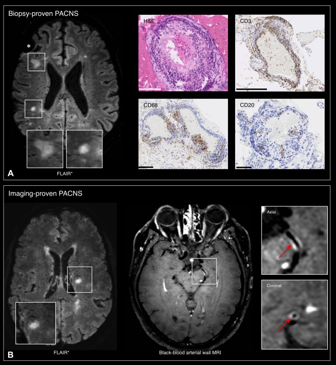
Figure 4.

Axial 3T FLAIR* images showing the presence of nonperivenular parenchymal lesions in 2 patients with primary angiitis of the central nervous system (PACNS). (A) Biopsy‐proven PACNS (57‐year‐old man; biopsy of the right frontal lobe and overlaying leptomeninges, asterisk). The histopathology shows the presence of a vasculocentric, transmural, multilayer inflammatory infiltrate (predominantly T lymphocytes) involving both the leptomeningeal and parenchymal arterioles. Scale bars: hematoxylin & eosin (H&E), 100 µm; CD3 (T lymphocytes), 250 µm; CD68 (macrophages), 100 µm; CD20 (B lymphocytes), 50 µm. (B) Imaging‐proven PACNS (48‐year‐old man). Vessel‐wall enhancement (arrows) of the left posterior and left middle cerebral artery is demonstrated using black‐blood arterial wall magnetic resonance imaging (MRI).
