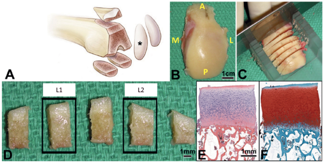
Figure 1.

Sample of lateral femoral condyle (black star) (right knee shown in example) was obtained (A, B). The orientation of the condyle was noted in the operating room (A = anterior; P = posterior; M = medial; L = lateral) and placed in an in-house fabricated miter box in AP orientation and cut into 4 mm thick arches through the region of the femur that is weight bearing in extension (C). The central region of 1 arch was cut into 5 specimens (4 mm x 4 mm), the second (Lateral) and fourth (Medial) were processed for histology (D). Each specimen was paraffin embedded, sectioned, and stained with HE (E), and SafO-FG (F) for analysis.
