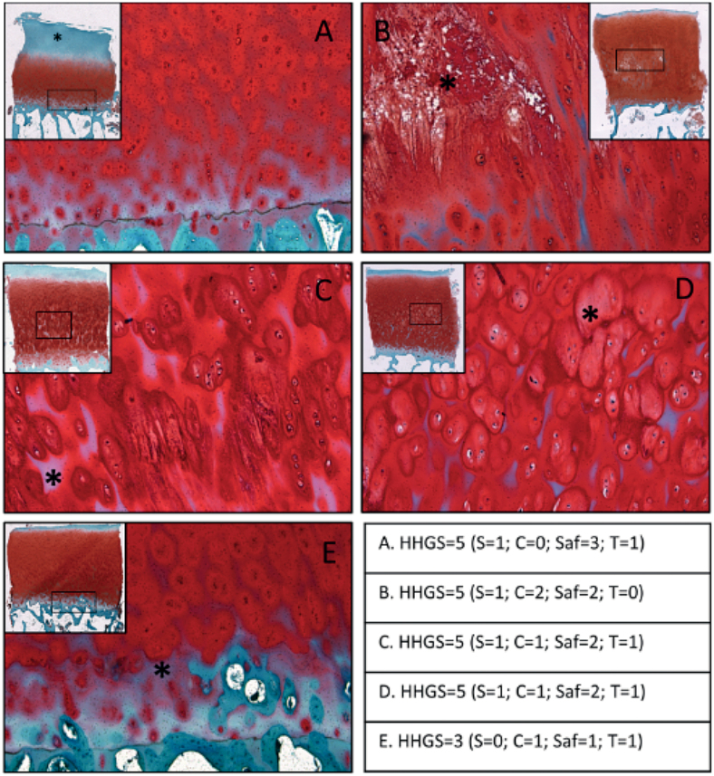
Figure 4.
Representative images of cartilage obtained from the lateral femoral condyle that indicate the unaccounted histopathological safraninO features: (A) loss of SafO stain in the top half of the tissue that is not associated with much surface erosion or fissures; (B) tissue necrosis/degradation in the radial zone, accompanied by some loss of SafO stain in the inter-territorial matrix region; (C) loss of SafO stain in the inter-territorial matrix, mainly confined to the bottom half of the tissue section; (D) varying staining patterns seen in the territorial matrix region and no SafO stain observed in some inter-territorial regions in the radial zone; (E) SafO staining loss near the tidemark even when the rest of the cartilage features appear relatively normal. The table indicates the total HHGS scores for each individual specimen, along with structure score (S), cell score (C), safraninO/fast green score (Saf), tidemark score (T). 43% of sample cohort presented with at least one feature.

