
Figure 10.
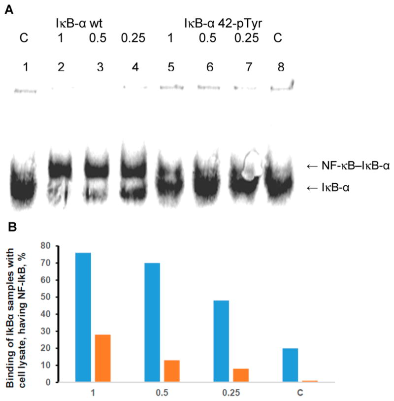
Binding of nonphosphorylated IκB-α (IκB-α wt) and phosphorylated IκB-α (IκB-α 42-pTyr) to NF-κB. (A) Analysis (native 8% polyacrylamide gel) of the purified lysate from activated Jurkat cells (Figure 8B, lane 5) in complex with 35S-labeled wild-type and phosphorylated IκB-α prepared by in vitro protein synthesis. Lanes 1 and 8, 35S-labeled protein not treated with lysate; lanes 2 and 5, 35S-labeled proteins treated with lysate; lanes 3 and 6, 35S-labeled proteins treated with lysate diluted 2-fold with 50 mM Tris–HCl, pH 7.4; lanes 4 and 7, 35S-labeled protein treated with lysate diluted 4-fold with 50 mM Tris–HCl, pH 7.4. (B) Quantification of the binding illustrated in the gel in panel A for 35S-labeled wild-type (blue) and phosphorylated (orange) IκB-α. C, control not treated with lysate; 1, purified lysate present at full strength; 0.5, lysate present at 2-fold dilution; 0.25, lysate present at 4-fold dilution.