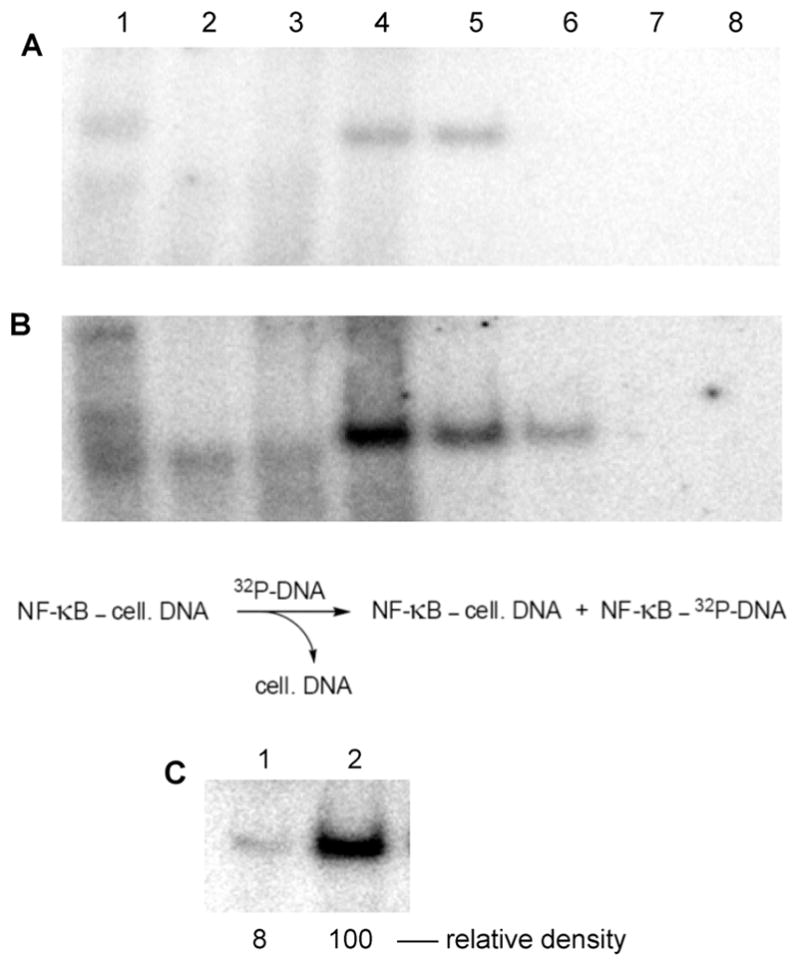
Figure 8.

Purification of NF-κB (as a complex with cellular DNA) from extracts of Jurkat cells (A) without initial cell activation and (B) following cell activation with PMA + A23187. The extracts were fractionated using DEAE-Sepharose CL-6B. Each fraction was incubated with 32P-labeled double-stranded DNA corresponding to the NF-κB binding site in the IL-2 gene promoter.8 Samples were separated by 6% native polyacrylamide gel electrophoresis and analyzed using a phosphorimager. Lane 1, crude extract; lane 2, flow through from DEAE-Sepharose CL-6B column; lanes 3–8, eluates resulting from wash with 100, 200, 300, 400, 500, and 600 mM NaCl, respectively. Formation of the NF-κB–32P-labeled DNA complex is envisioned by (partial) chemical exchange for cellular DNA. (C) Comparison of the amount of purified NF-κB isolated from unactivated vs activated Jurkat cells after purification by DEAE-Sepharose CL-6B (300 mM NaCl wash). Samples were separated by 6% native polyacrylamide gel electrophoresis and analyzed using a phosphorimager. Lane 1, purified NF-κB isolated from extracts of unactivated Jurkat cells; lane 2, purified NF-κB isolated from extracts of Jurkat cells activated with PMA + A23187.
