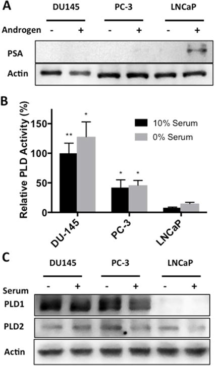
Fig. 1.

Phospholipase D activity is elevated in androgen insensitive prostate cancer cell lines. (A) DU145, PC-3 and LNCaP cells were plated at density of 200,000 cells/35 mm dish in complete media containing 10% charcoal stripped serum or 10% charcoal stripped serum supplemented with 10 nM testosterone. Cells were harvested and the levels of PSA were determined by Western blot analysis. (B) DU145, PC-3 and LNCaP cells were plated as in Fig 1A in complete media containing 10% serum or 0% serum overnight. Cells were then pre-labeled for 4 hr with [3H]-myristate followed by the addition of 0.8% 1-butanol for 20 min. PLD activity was determined by measuring the levels of the transphosphatidylation product phosphatidylbutanol as described in Materials and Methods. (C) DU145, PC-3 and LNCaP cells were plated as in Fig 1A in complete media containing 10% serum or 0% serum overnight. Cells were harvested and the levels of PLD1 and PLD2 were determined by Western blot analysis. No significance was determined for the difference in serum conditions within each cell line (p > 0.10). (*, p < 0.10; **, p < 0.05; ***, p < 0.01)
