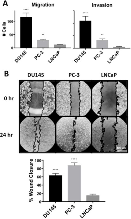
Fig. 3.

DU145 and PC-3 cells have higher migration and invasion than LNCaP cells. (A) DU145, PC-3 and LNCaP cells were placed in the upper chamber for a transwell migration assay at a density of 25,000 cells/6.5 mm transwell chamber in complete media containing 10% serum. Cells were allowed to migrate through the pores of the membrane to the lower chamber for 24 hr and were fixed, stained and scored under a microscope. Chambers with Matrigel™ indicated invasion while chambers without Matrigel™ indicated migration. (B) DU145, PC-3 and LNCaP cells were plated at a density of 100,000 cells/16 mm dish in complete media containing 10% serum. Cells were allowed to reach full confluence and a “wound gap” was scratched through the monolayer of cells. The media was changed to fresh media containing 10% serum and the “wound gap” was allowed to close for 24 hr. The area of closure was quantified using the MRI Wound Healing Tool macro for ImageJ. (**, p < 0.05, ****, p < 0.001)
