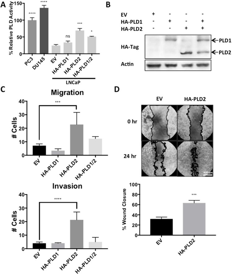
Fig. 5.

Overexpression of PLD2 in LNCaP cells increases migration and invasion. LNCaP cells were plated for transfection as in Fig. 4C with plasmids expressing empty vector or HA-PLD1 and/or HA-PLD2 as indicated. After 24 hours, cells were replated for PLD assay as in Fig. 1B (A), for Western blot analysis to determine levels of HA-tagged PLD1 and PLD2 (B), for migration and invasion transwell assay as in Fig. 3A (C), and for wound healing assay as in Fig. 3B (D). (ns, p > 0.10; *, p < 0.10, ***, p < 0.01, ****, p < 0.001)
