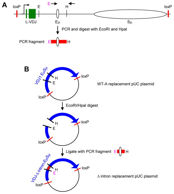
Fig. 2.
Schematic of the Δ intron replacement strategy. A) Map of WT-A replacement construct with loxP recombination sites (Baughn et al., 2011). Arrows represent the area amplified by PCR to generate the Δ intron fragment used for subcloning. The 5′ primer had a novel EcoRI (E) site (pink), and the 3′ primer was located near a HpaI (H) site. The PCR fragment containing the Eμ enhancer was then digested with E and H enzymes. B) Stepwise generation of Δ intron replacement plasmid. The WT-A replacement plasmid was cut with E and H, and ligated to the PCR fragment from A) to generate the Δ intron replacement plasmid.

