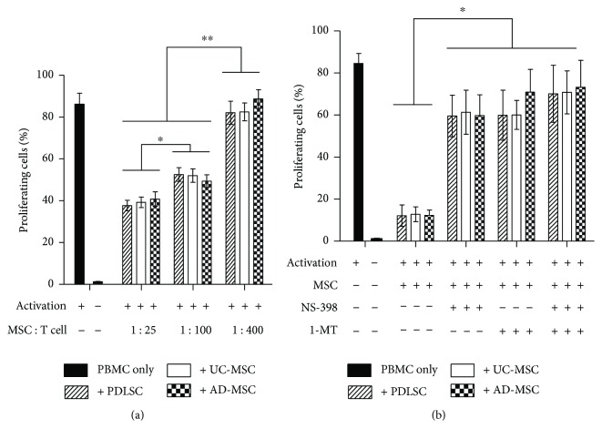
Figure 3.
(a) Dose-dependent inhibition of activated PBMC proliferation by MSCs. PBMCs isolated from healthy donors were labeled with CFSE, stimulated with anti-CD3/anti-CD28 antibody-coated beads, and cocultured with naive MSCs at MSC : PBMC ratios of 1 : 25, 1 : 100, or 1 : 400 for 3.5 days. Proliferating cells were analyzed by flow cytometry, and the percentage of each experimental group is represented as a bar on the histogram. (b) Recovery of PBMC proliferation by inhibitors of IDO or COX-2. Identical experiments were performed as those in (a) at an MSC : PBMC ratio of 1 : 10 in the presence of the IDO inhibitor 1-methyl-l-tryptophan (1-MT, 0.1 mM), the COX-2 inhibitor (NS-398, 0.1 mM), or both. Flow cytometric profiles are shown in Supplementary Figure 2. All experiments were performed independently at least three times. The graph shows the means and standard errors of the mean (SEMs). p values were obtained by ANOVA followed by Tukey's post hoc test. ∗ p < 0.05 and ∗∗ p < 0.01.

