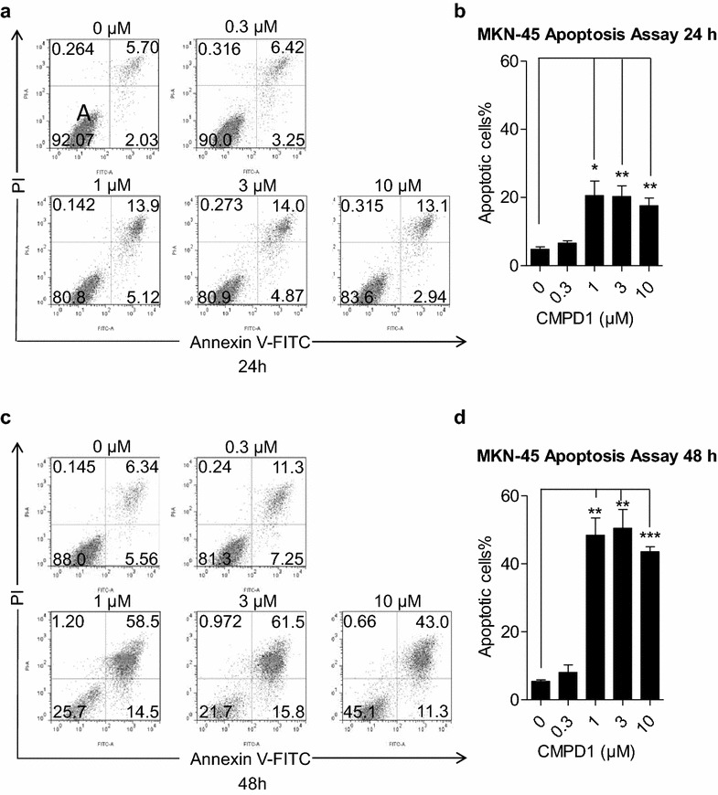
Fig. 2.

CMPD1 promoted apoptosis in MKN-45 cells. The upper-left, upper-right, lower-left, lower-right quadrants indicated necrotic, late apoptotic, viable and early apoptotic cell population, respectively. MKN-45 cells were treated with 0, 0.3, 1, 3 and 10 μM of CMPD1 respectively for a 24 h and c 48 h, and were then subjected to Annexin V-FITC/PI staining, followed by flow cytometer analysis. Quantification of the percentage of apoptotic cells treated with CMPD1 at various doses after treatment for b 24 h and d 48 h. *P < 0.05, **P < 0.01 and ***P < 0.001 vs. control
