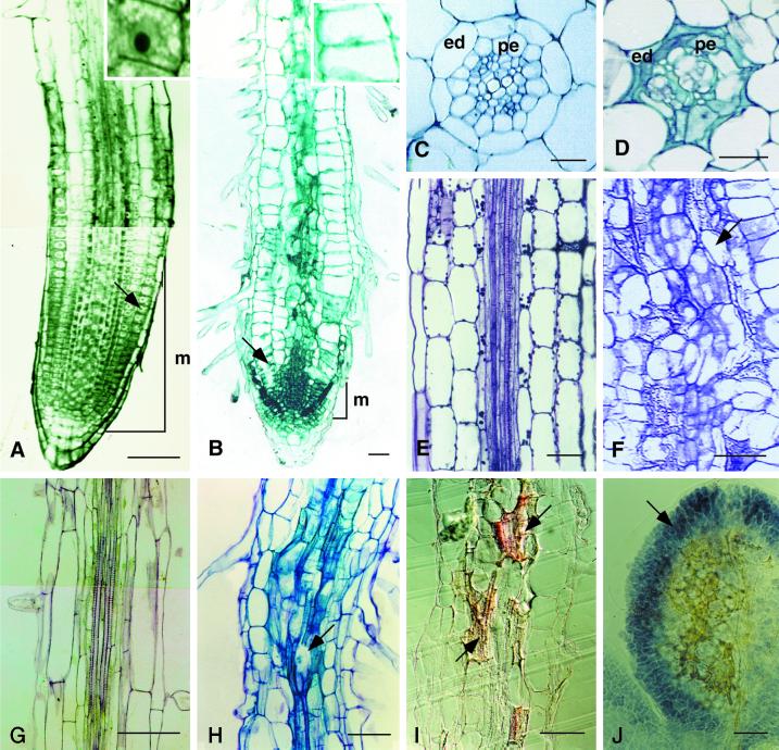
Figure 3.
Anatomy of the eld1 mutant and wild-type hypocotyls and roots. A, Longitudinal section of a 7-d-old wild-type root tip stained with TBO. The cell marked by an arrow is enlarged in the adjacent box. B, Longitudinal section of a 7-d-old eld1 mutant root tip was stained with TBO. The cell marked by an arrow is enlarged in the adjacent box. C, Cross-section of wild-type root showing diarch protoxylem in the stele surrounded by normal endodermis (ed) and pericycle (pe). D, Cross section of the eld1 root, showing disordered vessels, aberrant endodermis (ed) and pericycle (pe). E, Longitudinal section of wild-type hypocotyl showing parallel vascular cell files arranged tightly in the stele. F, Longitudinal section of the eld1 mutant hypocotyl showing a distorted vascular bundle and irregularly shaped vascular cells (marked by arrow). G, Longitudinal section of the upper region of the wild-type root, showing the stele. H, Longitudinal section of the upper region of the eld1 mutant root, showing abnormal vascular differentiation (marked by arrow). I, Longitudinal section of 7-d-old eld1 root stained with Sudan red 7B, suberin was stained red, and can be seen scattered randomly in the vascular bundle (marked by arrows). J, Cleared eld1 cotyledon stained with I-IK showing starch accumulated in the leaf periphery (marked by arrow) and suberized vein meshes in the middle. Bars = 100 μm (A and G–J), 50 μm (B, E, and F), and 20 μm (C and D).

