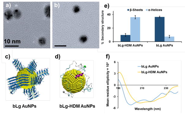
Figure 1.
Transmission electron microscopy (a, b) and discrete molecular simulations (c, d) show bLg AuNPs and bLg-HDM AuNPs. Circular dichroism spectroscopy indicates high β-sheet content in bLg AuNPs but not in bLg-HDM AuNPs (e, f). Scale bars in a, b: 10 nm. bLg amyloids of LACQCL (blue) coated on AuNPs (yellow spheres, 4 nm in diameter) (c). Full-length bLg molecules bound to an AuNP in the denatured state (d). Alpha-helices: purple, beta-sheets: orange, turns: cyan, coils: grey. Cα atoms in N- and C-termini: grey and green beads (d).

