
Fig. 7.
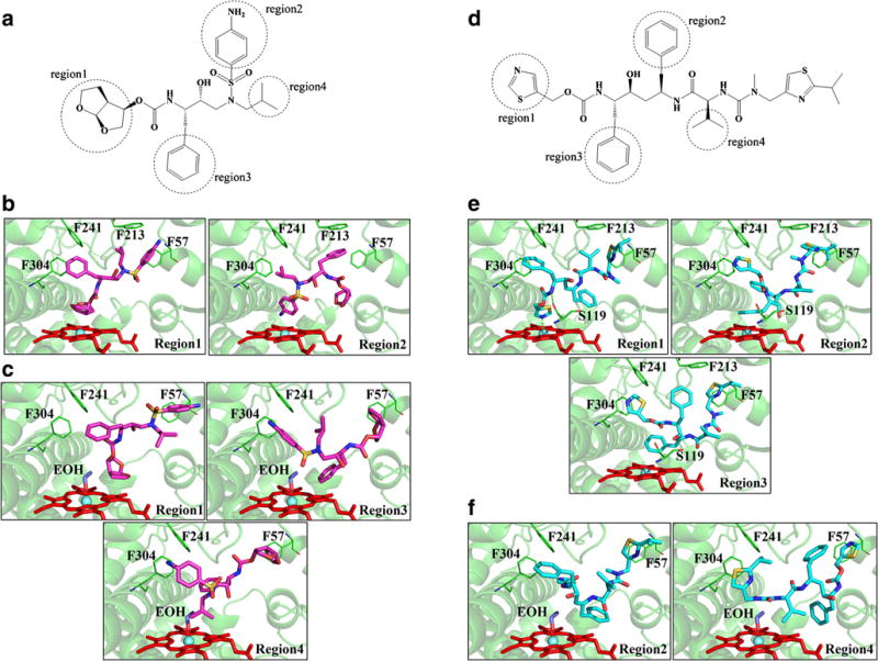
Molecular docking of Darunavir (DRV) and Ritonavir (RTV) with the human CYP3A4. (A) DRV (D) RTV interaction regions obtained through simulations. CYP3A4 binding sites and 3D model of DRV/RTV-CYP3A4 complex in absence (DRV (B), RTV (E)) and presence of ethanol (DRV(C), RTV (F)). All docking simulations were accomplished by GOLD suite 5.8. Residues within 20 Å of the ligand were defined as the binding pocket. Chemscore was used for scoring the ligand-CYP3A4 interactions. The first ten poses of scoring was catalogued as indicated regions in the figure.