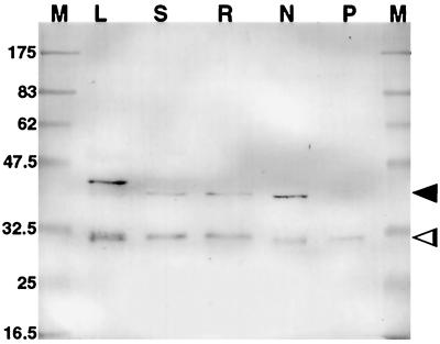
Figure 2.
Western blot of tissue extracts (10 μg of protein per lane) after electrophoresis using immunopurified R79 as the primary antibody to identify PsCYP15A antigen. M, Marker; L, leaf; S, shoot; R, uninoculated root; N, nodule; P, perisymbiont fluid. Two bands are apparent in all lanes except P, indicating pro- (arrowhead) and mature (open arrowhead) forms of PsCYP15A. Note the marginally slower mobility of the pro-form in the leaf sample. Molecular masses of marker proteins are represented in kD.

