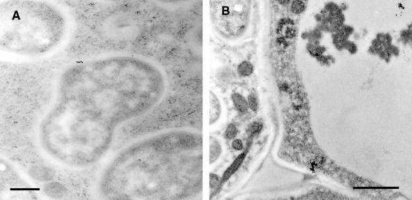
Figure 6.
Immunogold-labeled nodule sections treated with R79 antiserum and visualized by transmission electron microscopy. A, Cytoplasm of an infected cell. The center of the frame shows a complete symbiosome that has been labeled with 15-nm colloidal gold conjugate (indicated by the black dots), showing antigen localized within the space between the perisymbiont membrane and the bacteroid. Bar = 500 nm. B, Central tissue of a nodule, showing an infected cell (left side) and an uninfected cell (right side) separated by their respective cell walls. To the bottom of the frame, a further junction of these two cells with a third cell produces a large intercellular space. Both the cell walls and the intercellular space are unlabeled, whereas gold particles are associated with the dense material visible in the vacuole of the uninfected cell. Bar = 1 μm.

