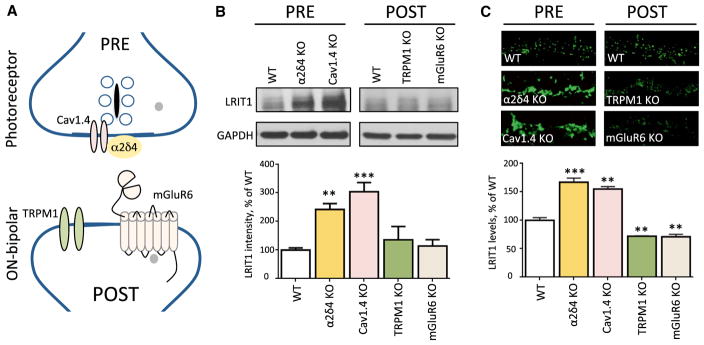
Figure 3. LRIT1 Synaptic Content Is Regulated by Changes in Presynaptic Release Apparatus.
(A) Scheme of the molecular organization of the photoreceptor synapse. Knockout mice lacking pre- and post-synaptic players depicted on the scheme were analyzed in the experiments.
(B) Analysis of LRIT1 protein expression by western blotting in total lysates prepared from retinas of the respective mouse strains. Retinas from 3–5 mice for each genotype were used for the quantification of the LRIT1 band intensities and the values were normalized to WT. **p < 0.01, ***p < 0.001; t test.
(C) Analysis of LRIT1 synaptic targeting by immunohistochemical staining of retina cross-sections of knockout mouse retinas as indicated. OPL regions are shown. Scale bar, 10 μm. The intensity of LRIT1 signal in the OPL was quantified and normalized to WT values. Two sections from each retina, two retinas per genotype; **p < 0.01, ***p < 0.001; t test.

