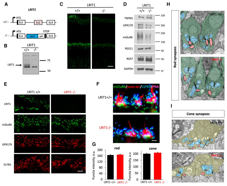
Figure 4. Generation and Characterization of Lrit1 Knockout Mice.
(A) Scheme for targeting Lrit1 gene. The deletion strategy included elimination of the critical coding exon 2 and introduction of the premature stop-codon preceding exon 3.
(B) Analysis of LRIT1 expression in wild-type and Lrit1 knockout (−/−) mouse retinas by western blotting.
(C) Analysis of LRIT1 localization in wild-type and Lrit1 knockout (−/−) mouse retinas by immunohistochemical staining of retina cross-sections, scale bar, 20 μm.
(D) Analysis of expression of proteins present in photoreceptor synapses by western blotting comparing wild-type and Lrit1 knockout (−/−) mouse retinas.
(E) Analysis of distribution of proteins present in photoreceptor synapses by immunohistochemical staining of retina cross-sections of wild-type and Lrit1 knockout (−/−) mouse retinas. OPL regions are shown, scale bar, 5 μm.
(F) Analysis of mGluR6 content in rod and cone synapses by immunohistochemistry. Staining with cone arrestin was used to define cone terminals and with PNA to identify active zones in the cone axons. Scale bar, 2.5 μm.
(G) Quantification of changes in mGluR6 staining in rod and cone synapses in the retinas of wild-type and Lrit1 knockout (−/−) mice.
(H) Analysis of rod synapse morphology by electron microscopy. Rod terminals are labeled in green, horizontal cell processes in blue and ON-bipolar dendrites in red.
(I) Analysis of cone synapse morphology by electron microscopy. Cone terminals are labeled in pale green, horizontal cell processes in blue and bipolar dendrites in red.

