
Fig. 2.
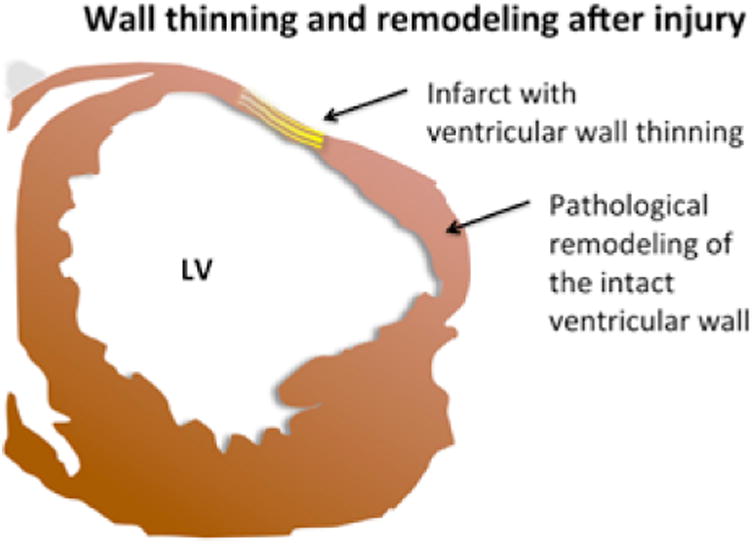
A graphical representation of a typical infarcted left ventricle (LV), showing wall thinning in the damaged area (yellow). Note that while not thinned, the wall adjacent can undergo pathological remodeling that can interfere with normal functioning.