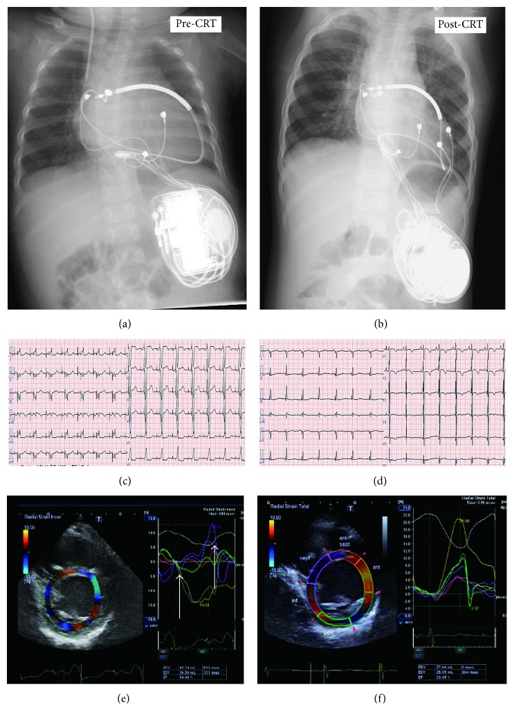
Figure 2.
Chest X-rays pre- and postcardiac resynchronization therapy (CRT). (a) The defibrillator was placed into the left upper abdomen, and the shock lead was fixed at the level of the superior vena cava. The right ventricular (RV) epicardial lead was placed at the RV free wall. (b) The left ventricular (LV) lead was placed at the LV lateral wall. The 12-lead ECG pre- and post-CRT. Before CRT (c), the QRS duration was 114 msec. After CRT (d), the QRS duration was shortened to 80 msec. Speckle-tracking radial dyssynchrony pre- and post-CRT (Artida™; Toshiba Medical Systems, Tokyo, Japan). Pre-CRT (e), dyssynchrony is shown as a time difference (arrow) between time to peak strain in the anterior wall (yellow) and septum peak strain (purple) (dual chamber (DDD) 90–150 bpm with an atrioventricular (AV) delay of 130 msec). Post-CRT (C-II), dyssynchrony disappeared (DDD 80–160 bpm, with an AV delay of 120 msec and a LV-RV delay of 0 msec).

